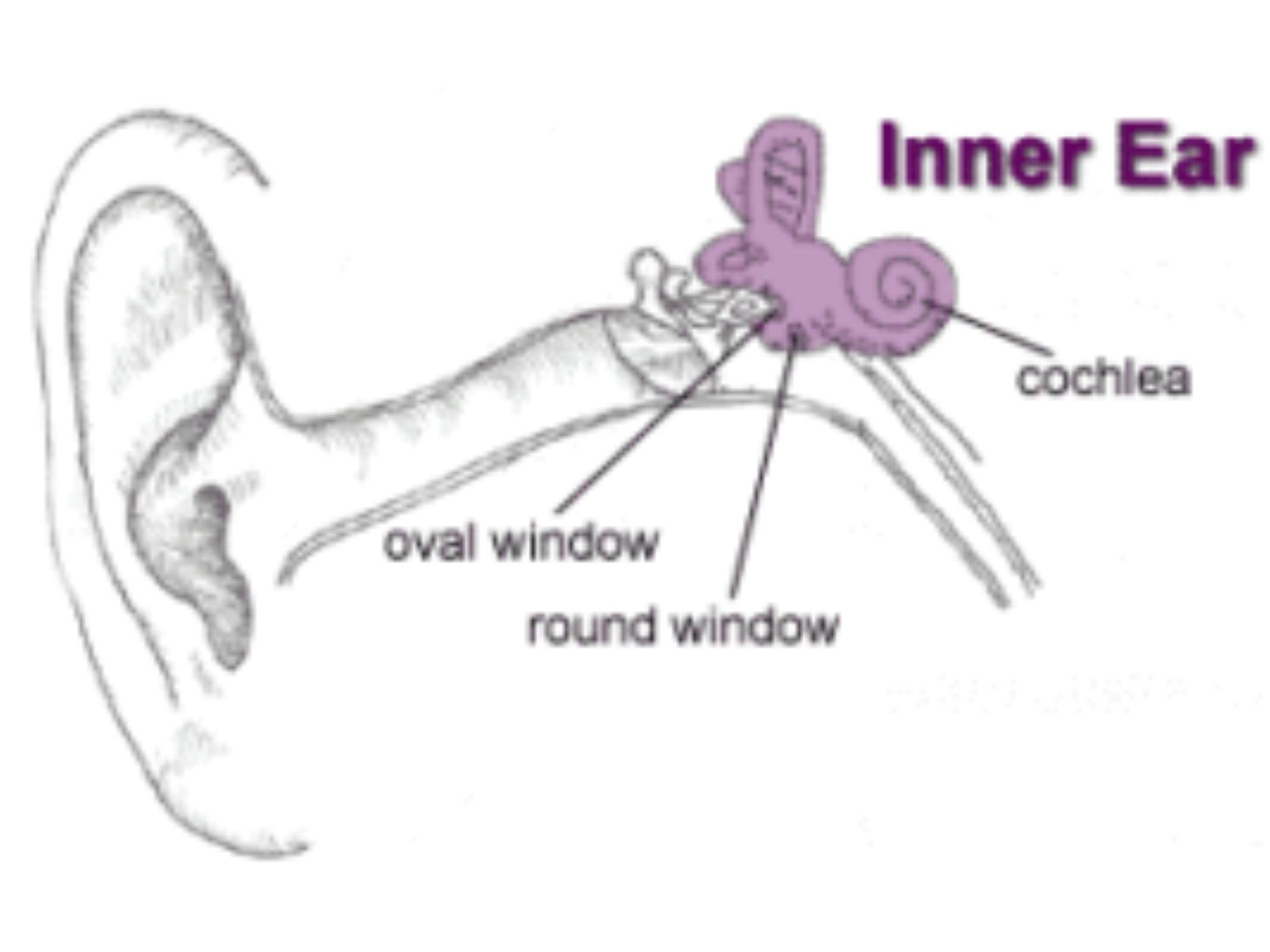
The Inner Part of the Ear
The purpose of the inner ear is to convert mechanical sound waves to neural impulses that can be recognized by the brain. The sensory receptors that are responsible for the initiation of neural impulses in the auditory nerve are contained in the cochlea of the inner ear. Our inner ear is the deepest part of your ear. The inner ear has two special jobs. It changes sound waves to electrical signals (nerve impulses). This allows the brain to hear and understand sounds. The inner ear is also important for balance.

The Cochlea
The Cochlea resembles a snail shell and spirals for about 2 3/4 turns around a bony column. Within the cochlea are three canals. They are called:
- The Scala Vesibuli
- The Scala Tympani (a bony shelf, called the spiral lamina, along with the basilar membrane and the spiral ligament, separate the upper scala vestibuli from the lower scala tympani)
- The Scala Media (cochlear duct)
The Scala Media
- The scala media is a triangular-shaped duct that contains the organ of hearing, called the “organ of Corti.”
- The basilar membrane, narrowest and stiffest near the oval window and widest at the tip of the cochlea, helps form the floor of the cochlear duct.
- The cochlear duct is separated from the scala vestibuli by Reissner’s membrane.
Hair Cells and Cilia
The surface of the basilar membrane contains phalangeal cells that support the critical hair cells of the organ of Corti. The hair cells are arranged with an inner row of about 3,500 hair cells and three to five rows of approximately 12,000 outer hair cells. The cilia of the hair cells extend along the entire length of the cochlear duct and are imbedded in the undersurface of the tectorial membrane. In general, the hair cells at the base of the cochlea respond to high-frequency sounds, while those at the apex respond to low-frequency sounds.
Activity in the Cochlea
The movement of the stapedial footplate in and out of the oval window moves the fluid in the scala vestibuli.
- This fluid pulse travels up the scala vestibuli but causes a downward shift of the cochlear duct, along with distortion of Reissner’s membrane and a displacement of the organ of Corti.
- The activity is then transferred through the basilar membrane to the scala tympani.
- At the end of the cochlea, the round window acts as a relief point and bulges outward when the oval window is pushed inward.
- The vibration of the basilar membrane causes a pull, or shearing force, of the hair cells against the tectorial membrane.
- This bending of the hair cells activates the neural endings so that sound is transformed into an electrochemical response.
- This response travels through the vestibulocochlear nerve and the brain interprets the signal as sound.
